QUOTE(dannyw @ Mar 29 2021, 01:19 PM)
Thanks for your reply.

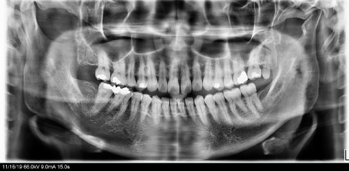
1- You able to detect the periodontitis from the xray? So far, I been scaling every one to two years.
2- How to identify the generalised loss of bone between teeth? None of my dentist mention any about this.
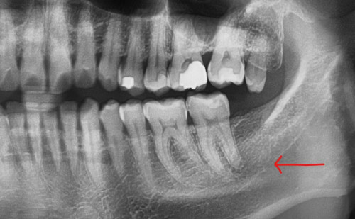
3-a- I have get the second advice from another dentist, which commented the same. It could be developed secondary caries based on the horizontal line from side. That could be during my extraction of my wisdom teeth. But my wisdom teeth been surgery out more than 20 years. I’m not sure still possible?
b- Anything I can do to improve it? I did notice every time I floss there is some small hole stuck my floss. It located quite deep, nothing can do about it.
c- I show this xray to the dentist, he did say the same, not really clear and hard to judge. However, he managed to see the borders should be still well.
He did say the same on the vitality test, just before this, I still have feeling when touch or even with the cold air blow.
So his conclusion is monitor it, unless I have chewing problem or pain then only go for further checking.
After that recent filled, it still have some discomfort on and off, not pain level. And is improving this few days. Hope it will go off. So worry on the RCT.

oh ya, if worst scenario happened, can it be just extract rather than go through the tough and not guarantee process?
Anyway, really appreciate your time and advice.
1- Yes, its very clear because of the bone loss levels shown in the xray. Scaling is not itself a solution, you need to improve your oral hygiene. You need to change your brushing technique, learn to use floss and include use of mouthwash. In the beginning Dentist might advise you a medicated toothpaste. Scaling is a critical part of treatment regime, but its effective in combination.
2- I can not say why your dentists didnt advise Oral Hygiene Instructions to you, periodontitis is as clear as day light here. You might also be suffering from bleeding gums and develop bad breath from time to time. If you are a smoker, cut down on number of cigarettes.
3- a-The 2nd opinion dentist was stating that since there is no 3rd molar, the caries could have developed from the far side, again if that is the case the reason is your periodontitis. In healthy gums, the root material is not exposed instead covered by a combination of bone and gums. The material on the root is not as resistant to caries as the one on the top of tooth. I simplified tooth anatomy for you, the interaction is far complex.
b- Yes, ask your dentist on how to properly brush your teeth, and the frequency of brushing. You might need scaling and root planning (a special type of scaling) Refer to point 1.
c- Vitality test consists of hot and cold test, some times electrical stimulus is also used. If your tooth is sensitive to touch, aka Tender to touch, further probing is required to establish the cause for it.
I do not know what discomfort means here, its a very subjective term. There shouldn't be any pain or tenderness, if so antibiotics might be required to control the infection.
Once RCT is performed, the tooth must be crowned as once the pulp is removed tooth becomes very brittle and could crack. Extraction should be the last option, when everything else fails. In your case you need to improve oral hygiene.
Again I wont comment on dentists competency, but if they keep on missing developing and worsening of your gum disease, go figure.
This post has been edited by a-y: Mar 29 2021, 06:37 PM 

 Mar 18 2021, 04:45 PM, updated 5y ago


 Quote
Quote

 0.0153sec
 0.47
0.47
 5 queries
 GZIP Disabled